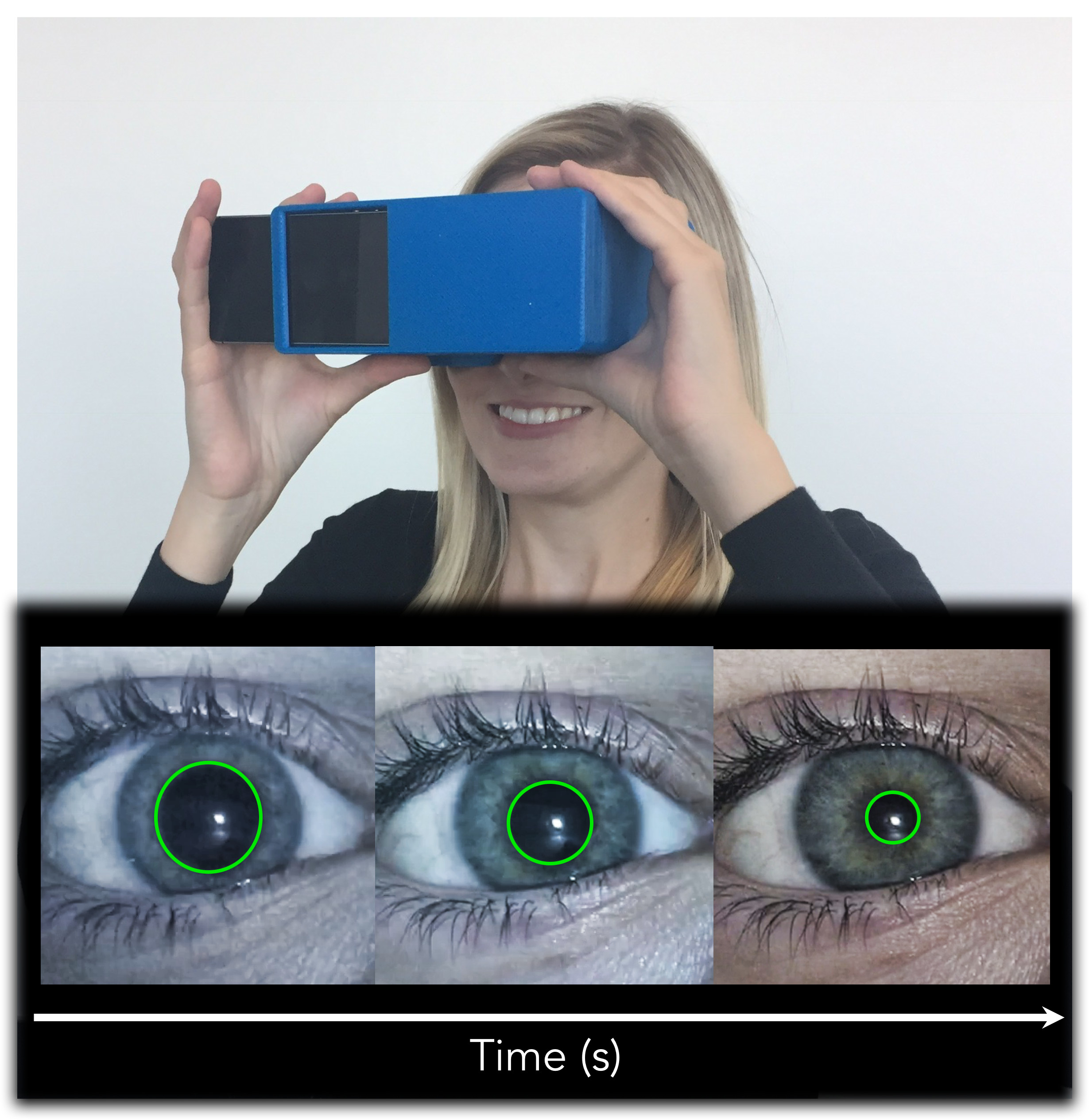

Once again, this is by no means an exhaustive list of assessment tools available. similar to looking out a car’s side window while moving)ĥ) Electronystagmography (ENG)– advanced electrical testing of various ocular capabilities Insight into the workings of the ocular (eye) system and its connections with most parts of the brain.ġ) Vestibulo-ocular reflex (VOR) testing – often performed with the patient focusing on an object while the examiner moves the head, or while the patient is rotated in a chairĢ) Testing for eye saccades (fast movements between targets) – movements are typically over or under-compensated with a concussionģ) Cortical blind spot mapping – mapping of the visual defect created by the optic nerve attaching into your retina where there are no receptors for light (rods/cones)Ĥ) Testing for optokinetic nystagmus (OKN) – reflexive eye movement caused by tracking of movement within a visual field (i.e. That said, there are a host of other very sensitive tests that can offer clinicians incredibly valuable. As well as the higher brain centers that regulate these functions. And can reveal a great deal about the integrity of these cranial nerves. While not an exhaustive list, these tests can be done in a very short period of time (5 minutes or less). Well, I’m here to tell you that they need to be looking at a great deal more than that! Some basic tests to look for when you or a loved one is examining for suspected concussion are as follows: 1) Observation for eye malposition 2) Direct and indirect pupil response to light (as noted above) 3) Cardinal fields of gaze (eye movements in all directions) 4) Observation of slow and fast eye movements, and termination of said movements 5) Eye convergence (crossing of the eyes – as your mother told you to never do!) 6) Ophthalmoscopic examination (looking inside the eyes) 7) Visual acuity/Snellen chart (how well you see) 8) Eye cover/uncover testing (more sensitive test for eye deviation) 9) Eye dominance and/or suppression testing As the one and only diagnostic factor for concussion. A doctor shines a light in someone’s eyes to look for a lack of pupil constriction.
#Concussion pupil test tv
We have all seen it at some point on TV or the ‘Big Screen’. In recent years that did not have some type of visual or oculomotor (eye movement) consequence. I cannot honestly recall a single case of concussion presenting to my office. Light sensitivity-photophobia, eye fatigue, double vision-diplopia, reading difficulties, etc.).

Shifting our focus back to the eyes (pun intended) most individuals that have suffered a concussion will complain of some type of symptom related to eye function (e.g. The longer one’s brain is adapting to negative changes incurred as a result of a head injury (referred to as maladaptive plasticity), the longer it will take to rehabilitate their way out of them!

This is entirely unacceptable as early intervention is critical with concussions, as is the case with most disorders of humankind, and it may significantly decrease the likelihood of more serious consequences. Patients are often treating with a ‘sit-and-wait’ approach meaning it is only after signs and symptoms have manifested and worsened that people often seek the care of their own accord. Given concussions are ascribing to this ‘low severity’ status, timely evaluation and treatment are often poor or non-existent at best. Most experts would agree that these are the least serious type of brain injury yet left untreated many can suffer devastating and debilitating consequences such as vertigo/dizziness, balance problems, cognitive dysfunction, emotional disorders, headaches, and many others. A concussion comes from the Latin ‘Concutere’, which means to shake violently. There are many types and causes of head injury, but, by far, the most common are concussions related to motor vehicle accidents and contact sports. It is also critical to assess higher brain centers that control various eye reflexes, which will be discussed later in this article, during the eye examination as well. Of these cranial nerves, the most telling findings will likely come from the ones involved in vision and eye movement (cranial nerves 2, 3, 4, and 6).
#Concussion pupil test professional
While a thorough neurological history and examination with a qualified professional should be performed for any suspected head injury, particular emphasis should be placed on the cranial nerves – the nerves that exit your brain and brainstem.


 0 kommentar(er)
